Corneal Topography Test
Published on October 27th, 2025
 Canada

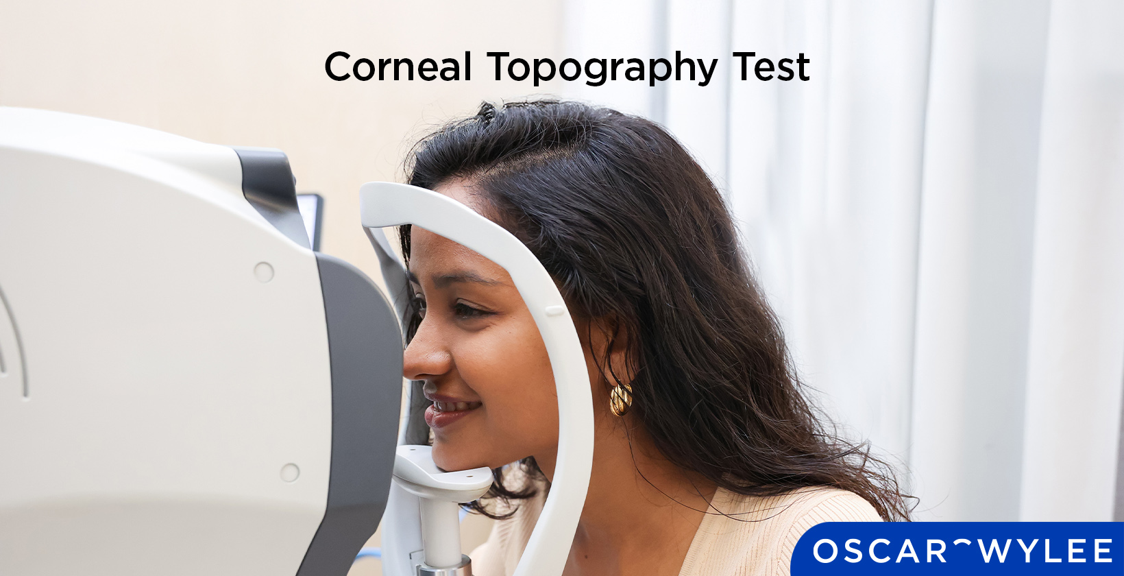
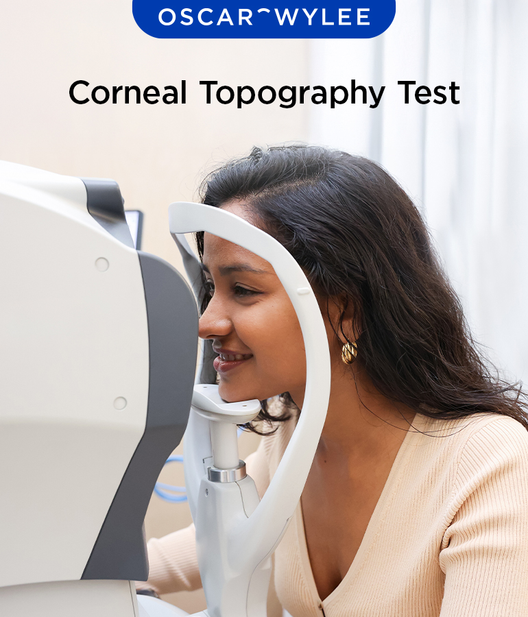
A Corneal Topography Test provides a two-dimensional map of the cornea's shape and curvature in order to allow for the detection of corneal diseases. The test will display a colour-coded image of your corneal shape and can be conducted by optometrists or ophthalmologists. It is important to note that Oscar Wylee does not conduct Corneal Topography Tests. Read on to learn more about Corneal Topography tests, including the conditions they can detect and what to expect.
What is Corneal Topography?
A Corneal Topography is a non-invasive eye exam undertaken to measure and map an eye's cornea by taking two-dimensional and cross-sectional images of it. The cornea refers to the clear outer layer at the front of the eye, which helps your eye to focus light so you can see clearly. It can be an important test to take for processes such as screening for refractive surgery, contact lens fitting, and planning arcuate incisions for astigmatism correction, according to the National Library of Medicine.
How Does a Corneal Topography Test Work?
The corneal topography works by producing a series of maps that display the surface features of your cornea. These are displayed through various colours and numbers. The test can produce many different types of maps, depending on what the ophthalmologist needs to learn about your eye health. The most common maps include the Axial map showing the cornea curvature, the Elevation map showing the outward curvature and the Corneal thickness map.
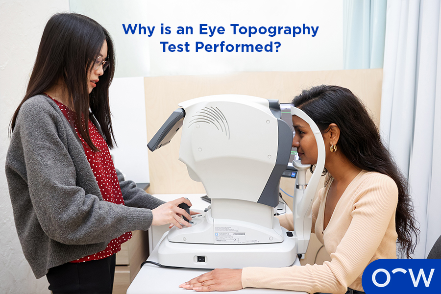
Why is an Eye Topography Test Performed?
An eye topography test can be performed for a myriad of reasons, such as evaluating corneal deformities or injuries, planning surgery for refractive errors, treating and evaluating astigmatism and specialised contact lens fits. Doctors and optometrists will recommend it as it can detect these eye diseases with high accuracy.
What Eye Conditions Can Corneal Topography Detect?
The eye conditions that corneal topography can detect include the detection of eye growths, astigmatism and keratoconus. Read on for more information on these eye conditions that can be detected by corneal topography below.
- Detection of eye growths: The detection of eye growths can be done through corneal topography, as it detects growths such as pterygium. It is a raised, fleshy growth on your eye's conjunctiva, which may look whitish or pinkish.
- Astigmatism: Astigmatism refers to when the shape of your eye is more curved than it should be, leading to blurred vision. Astigmatism is usually caused by your cornea having an irregular shape, called corneal astigmatism. There is also lenticular astigmatism, where the lens in your eye has an irregular shape, often caused by cataracts.
- Keratoconus: Keratoconus is an eye condition wherein the normally rounded cornea bulges outward into a cone shape. Keratoconus can lead to irregular astigmatism when the cornea changes to a cone shape, or nearsightedness if the front of the cornea steepens.
What do Normal and Abnormal Corneal Topography Results Mean?
A normal corneal topography will display predominantly more cool colours like green and blue, which show flatter areas of the cornea. Normal corneal topography on an axial map, which shows the curvature of your cornea, is mostly green. An abnormal corneal topography will show more warm colours like red and orange predominantly, indicating a steep cornea that can point to an eye condition present, such as astigmatism.
How is Corneal Topography Used for Astigmatism and Keratoconus?
Corneal topography is used for the diagnosis and monitoring of astigmatism and keratoconus. It is invaluable for early detection of these conditions, even before the emergence of symptoms.
How is Corneal Topography Used in LASIK, PRK, and Refractive Surgery?
Corneal topography is used in LASIK, PRK and other refractive surgeries as a pre-surgery screening. This is to determine whether to perform surgery on highly irregular and asymmetrical astigmatism according to the British Journal of Ophthalmology. Both LASIK and PRK aim to reshape the cornea to correct astigmatism.
How does Corneal Topography Help with Contact Lens Fitting?
Corneal topography can help with contact lens fitting, as abnormalities in the cornea, such as a highly steep cornea, may require custom contact lenses for optimal fit and comfort. Characteristics of the cornea, such as the central curvature, symmetry, and asphericity, also contribute to the fit of contact lenses.
What is the Difference Between Corneal Topography and Corneal Tomography?
The difference between corneal topography and corneal tomography is the parts of the eye they capture. Corneal topography is used to provide a two-dimensional view of the corneal surface in order to detect corneal diseases. Corneal tomography uses light waves to capture high-resolution images of the cornea, iris, and anterior chamber according to Ento Key.
What is Keratoconus Topography?
Keratoconus topography refers to the characteristics of the topography results that emerge as a result of Keratoconus. Keratoconus is a disease that results in thinning of the central zone of the cornea, according to McGill University. A corneal topography will show the affected cornea as having a cone-shaped appearance.
What do Corneal Topography Images and Eye Maps Show?
Corneal topography images and eye maps show the surface curvature and shape of the front of the cornea. This is visually displayed using concentric rings, where widely spaced rings indicate a flatter cornea, and closely spaced rings indicate a steep cornea.
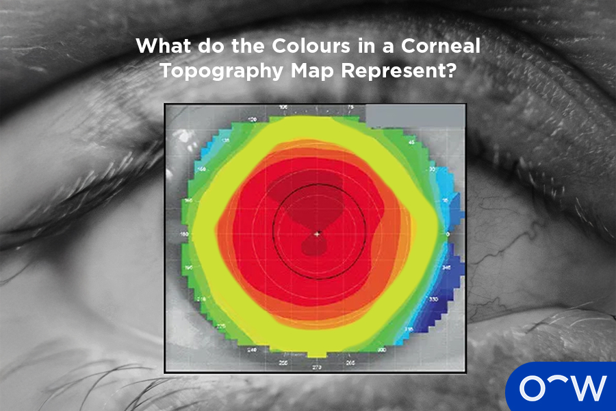
What do the Colours in a Corneal Topography Map Represent?
The colours in a corneal topography map represent the characteristics of the cornea. The warm colours, such as red, orange and yellow, represent the steepness of the cornea, while the cool colours represent how flat the cornea is. These colours also indicate cornea thickness, as warm colours indicate where the cornea is thinner, while cool colours indicate where in the cornea it is thicker.
How Accurate is the Corneal Topography Test?
The Corneal Topography Test is highly accurate and a legitimate test used by Ophthalmologists and other eye care professionals to create a three-dimensional map of the cornea. It is highly accurate in diagnosing eye diseases such as Keratoconus, with a 96% accuracy rate according to Ento Key. It is important to note that Oscar Wylee does not conduct Corneal Topography Tests, and an Ophthalmologist should be consulted for this procedure.
What Should You Expect During a Corneal Topography Eye Exam?
You should expect a quick and unobtrusive process during a corneal topography eye exam. You will be simply asked to rest your head against a bar and sit in front of a lighted bowl that contains a pattern of rings. A series of data points will be collected, and a colour-coded image of your corneal shape will be generated on a computer screen for the ophthalmologist to assess.
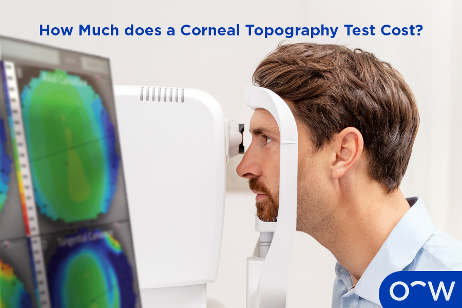
How Much does a Corneal Topography Test Cost?
The cost of a corneal topography test will depend on the Ophthalmologist who conducts it. The price range can differ between different clinics, and therefore, it may not be accurate to list just one figure.
Where Can You Get a Corneal Topography Test Near Me?
You can get a corneal topography test done near you by searching online. Simply type into Google 'Corneal Topography near me' and search results will appear for locations where they can be undertaken. They are often available at Ophthalmologists and some Optometrists. It should be noted that Oscar Wylee does not do Corneal Topography Tests.
Read Corneal Topography Test in other Oscar Wylee regions and their languages.
Canada